
Tracheal wash from a horse
Case Information
A 22 year old Standardbred mare was admitted to the Cornell Large Animal Medicine Service for smoke inhalation after a barn fire where 26 other horses died. At presentation (6 hours after the initial insult), the mare was anxious and dyspneic, with burns on the face and neck (including the cornea as seen byhyperemic conjunctiva and edema of eyelids) and a singed mane. Thoracic auscultation revealed severe bilateral crackles and harsh lung sounds, with increased respiratory effort. Foam was present at the nares and mouth. Initial bloodwork revealed a relative increase in hematocrit (52%, reference interval, 31-47%) due to hemoconcentration, and a stress and inflammatory leukogram characterized by a moderate leukocytosis (24.0 x 10³/µL, reference interval, 5.5 – 11.4 x 10³/µL) attributable to a mature neutrophilia and a mild to moderate lymphopenia (0.7, reference interval, 1.6 – 6.6 x 10³/µL). Inflammation was supported by high fibrinogen concentration (500 mg/dL, reference interval, 0 – 200 mg/dL) determined by heat precipitation. Total protein (6.5 g/dL, reference interval, 5.2 – 7.8 g/dL) and serum albumin (2.8 g/dL, reference interval, 2.8 – 3.8 g/dL) were both within reference intervals, but were considered to be low in light of the hemoconcentration. Whole blood arterial gasometry revealed a respiratory alkalosis associated with hyperventilation.
Hours after initial stabilization, the mare relapsed into severe dyspnea; endoscopic examination revealed occlusion of upper airways due to severe edema. A tracheostomy and placement of a tracheal tube were performed. Samples of a transtracheal aspirate were submitted for cytologic evaluation. Representative cytologic images are provided below. After examining the images, answer the following questions.
- Is there evidence of inflammation? If so, how would you classify it?
- Provide two possibilities for the black granular material present within macrophages. How can you distinguish these?
- Identify the arrowed structure in Figure 3.
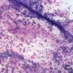
Figure 1: Tracheal wash direct smear (Wright’s stain 100x) 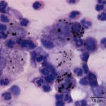
Figure 2. Tracheal wash direct smear (Wright’s stain 1000x) 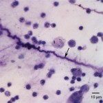
Figure 3. Tracheal wash direct smear (Wright’s stain 500x)
Answer on next page