Peritoneal fluid from a horse
Case information
A 5 year old Thoroughbred-paint gelding presented to the Cornell University Large Animal Hospital with a history of increased respiratory effort, abdominal distention, and depression after turn out to a new pasture. On presentation, the horse was quiet, alert and responsive, and vital signs were within normal limits. Physical examination revealed abdominal distention and pain, decreased intestinal sounds, and abnormal rectal palpation (absence of fecal material, distended cecum, and dilated loops of small intestine). Due to the marked abdominal distention, the cecum was trocharized and some of the gas distention was relieved. The horse was treated with intravenous fluids and 2% lidocaine (250 mL as a constant rate infusion) for pain control and monitored overnight. The day after initial presentation, the horse’s condition had improved and rectal examination revealed normal formed manure and decreased colon distention. The lidocaine infusion was discontinued. On the second day of hospitalization, blood and peritoneal fluid were collected and submitted for a complete blood count and biochemical analysis, and cytologic evaluation, respectively.
Blood work revealed a mild non-regenerative anemia with a hematocrit of 32% (reference interval [RI], 34-46%) interpreted as decreased erythropoiesis in response to inflammation. There was an inflammatory leukogram due to the presence of a mild left shift (0.3 thousand/µL; RI, 0.0-0.1 thousand/µL), and mild toxic change, despite normal total leukocyte (5.5 thousand/µL; RI, 5.2-10.1 thousand/µL) and neutrophil (4.6 thousand/µL; RI, 2.4-6.6 thousand/µL) counts. There was a mild lymphopenia of 0.6 thousand/µL (RI, 1.2-4.9 thousand/µL) supporting concurrent stress (glucocorticoid response). Biochemical results were overall unremarkable.
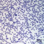
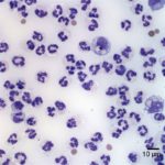
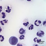
The submitted peritoneal fluid was light-orange in color and opaque, with a total protein by refractometry of 5.5 g/dL (upper reference limit, <2.5 g/dL). The total nucleated cell count was 1004.6 thousand/µL (RI, <5.0 thousand/µL) and the red blood cell count was 314.2 thousand/µL. Direct smears from the submitted fluid were prepared and examined.
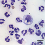
Evaluate the representative photomicrographs of the peritoneal fluid shown below and answer the following questions:
- Identify the cells labeled as “A” and “B” in Figure 3.
- How would you classify the effusion? Can you identify a cause?
- What do you think the prognosis would be for this horse?
|
|
|
|
|