
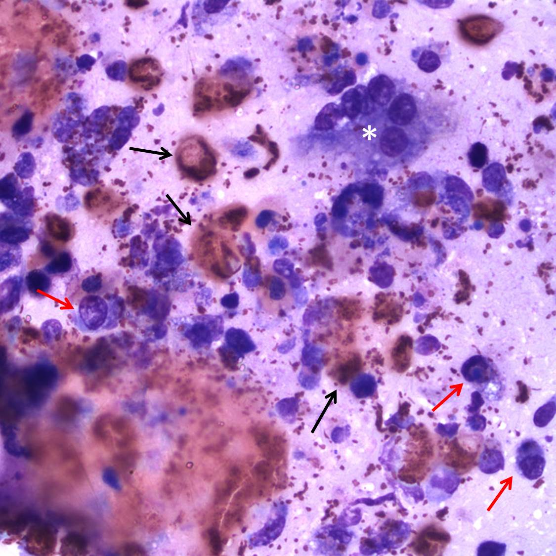
Figure 2B. Another view of the mixed inflammatory cells features a multinucleated giant cell (white asterisk). Note the thick aggregate of heterophils in the bottom left corner of the figure. A few individual heterophils and macrophages are identified (black and red arrows, respectively). Wright’s stain, 500x.