
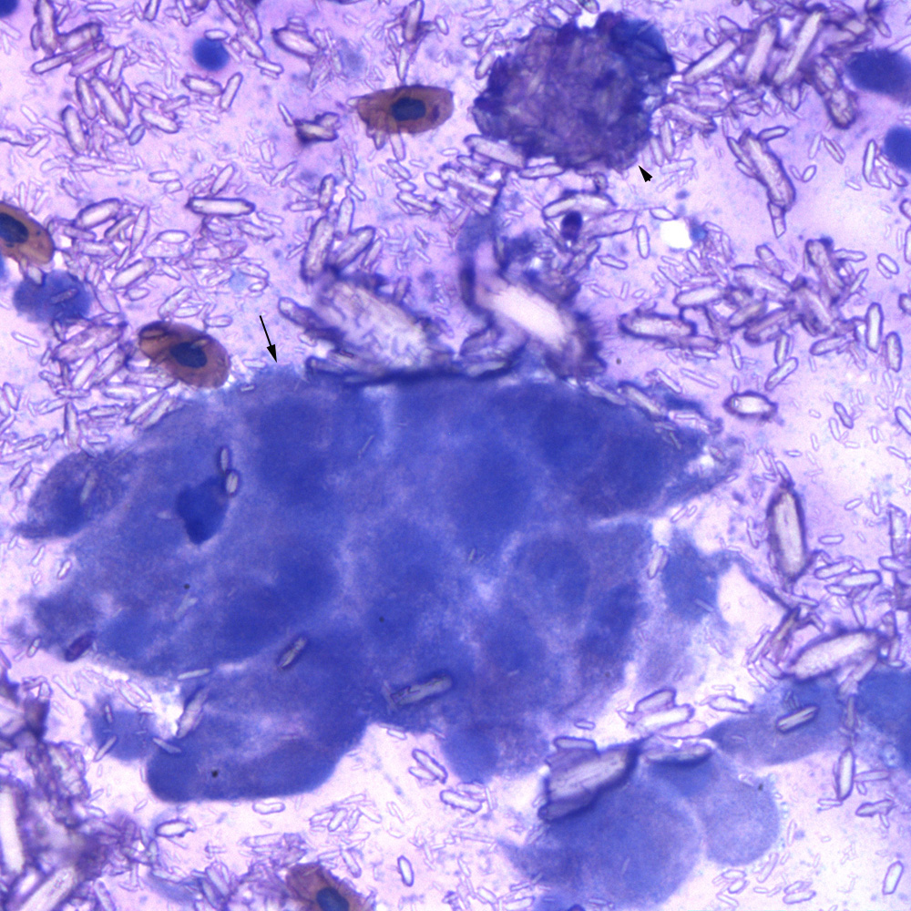
A higher power view shows that the epithelial cells are round to polygonal with distinct cell boundaries (arrow). There are numerous extracellular largely parallel-sided colorless to light pink crystals of variable size and thickness. The macrophage contains many phagocytized crystals in the cytoplasm (arrowhead). There are also low numbers of erythrocytes, which are the likely source of the pigment (presumptive, hemosiderin), noted in macrophages on histologic evaluation of the mass (although erythrophagia was not definitively observed in the cytologic samples) (modified Wright’s stain, 50x objective).