
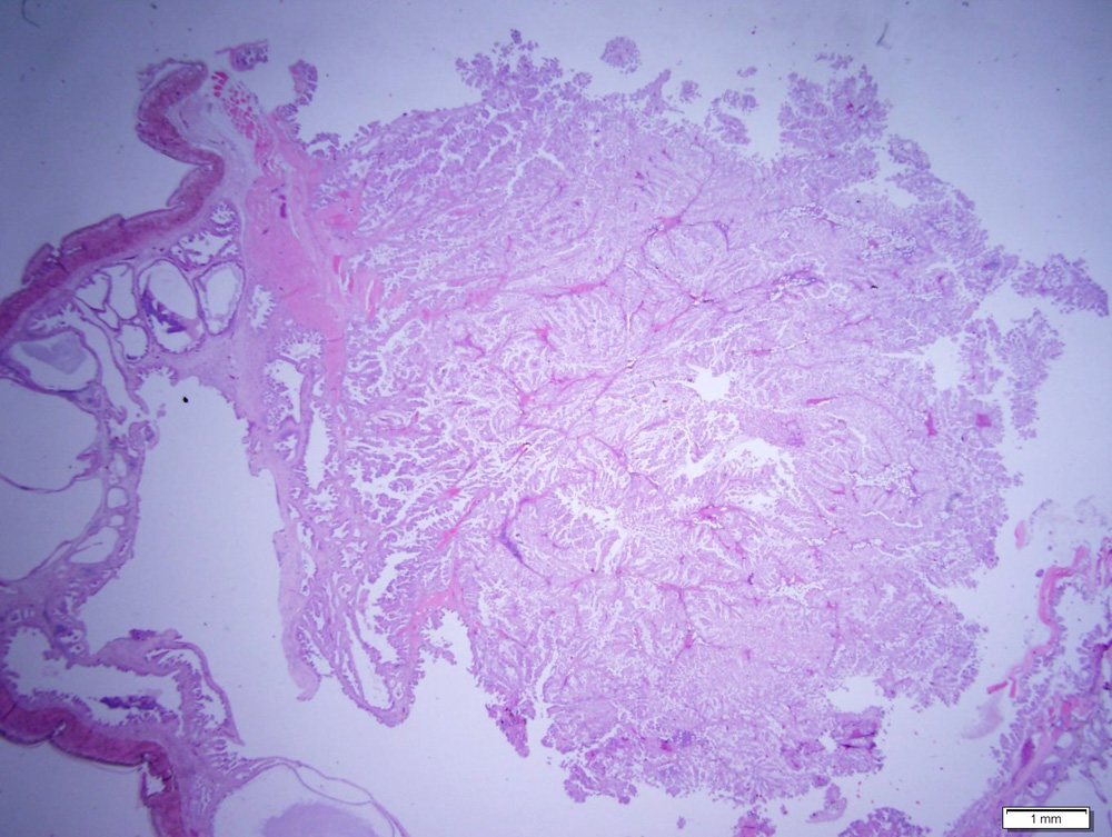
In this lower power image of the smaller mass, the mass is arising off the wall of the endolymphatic sac (the lining of the sac is seen to the left of the image). The mass consists of papilliform arrangements of columnar epithelial cells, supporting by fine fibrovascular stroma. In some areas of the mass (lower and upper right hand side), the epithelial cells are more disorganized and forming dense lobules or clusters and have lost their single cell lining arrangement (H&E stain, 1.25x).