
Peripheral blood and pleural fluid from a dog
Case information
A 6.5 year old female spayed Cavalier King Charles Spaniel presented to the Cornell University Emergency Service for workup of an increased peripheral white blood cell count and pleural effusion detected by the primary veterinarian. Prior to presentation at Cornell, the primary veterinarian had removed approximately 300 mL of fluid from both sides of the chest. At presentation, the dog was quiet, alert, responsive, and tachypneic (44 bpm) with shallow breaths. Lung sounds were slightly decreased ventrally, compatible with an effusion. Thoracic radiographs confirmed a pleural effusion and revealed deviation of the trachea to the right with an enlarged mediastinal mass. The dog was placed into an oxygen cage due to low blood oxygen saturation (94%) on arterial blood-gas analysis and given intravenous fluid therapy at 1.5 times maintenance. Blood and pleural fluid were collected before treatment. Results are shown on the tables below.
| Pertinent hematologic results | |||
| Test | Result | Units | Reference interval |
| Hematocrit | 40 | % | 41-58 |
| Retic count | 0.4 | % | 0.2-1.5 |
| Absolute retic count | 18.9 | thou/μL | 11.0-92.0 |
| White blood cells | 227.6 | thou/μL | 5.7-14.2 |
| Seg neutrophils | 4.6 | thou/μL | 2.7-9.4 |
| Band neutrophils | 2.3 | thou/μL | 0-0.1 |
| Other leukocytes | 211.7 | thou/μL | 0-0.1 |
| Platelet count | 108 | thou/μL | 186-545 |
| MPV | 17.5 | fL | 8.4-14.1 |
| Mild toxic change in neutrophils | |||
| Pleural fluid | ||
| Test | Result | Units |
| Volume | 1.8 | mL |
| Color | Medium red | |
| Turbidity | Opaque | |
| Total Protein (ref) | <2.5 | g/dL |
| Nucleated cells | 16.0 | thou/μL |
| Red blood cells | 178.1 | thou/μL |
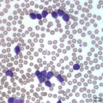
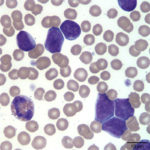
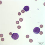
The “other cells” are shown on the representative photomicrographs of the blood and pleural fluid below. Based on the provided data, answer the following questions:
- What is the best interpretation for the leukocytosis?
- What additional diagnostic tests would you recommend for classification of the type of neoplasm?
|
|
|
|
Answers on next page