
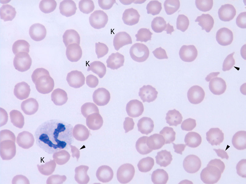
Numerous keratocytes (K) and schistocytes (arrowhead) are seen in this dog with disseminated intravascular coagulation secondary to acute hepatic necrosis. There are also some target cells, which are likely secondary to lipid alterations in the RBC membrane from the liver disease. A segmented neutrophil displaying mild toxic change is also present but no platelets are identified (thrombocytopenia is a characteristic feature of overt or fulminant DIC in dogs) (Wright’s stain, 1000x magnification).