
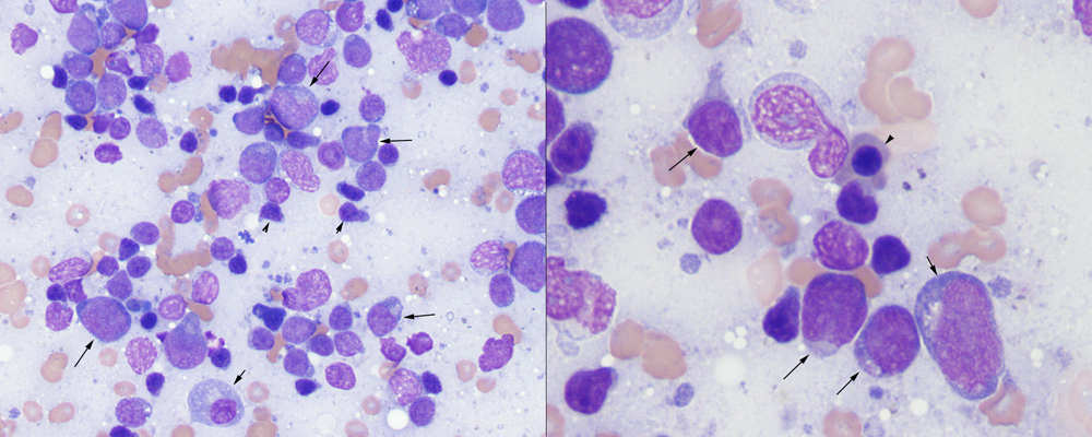
Acute myeloid leukemia in a lymph node aspirate from a dog. Left panel: Note the infiltrative blasts (arrows) in among residual normal lymph node constituents, including a plasma cell (short arrow) and numerous small lymphocytes (arrowheads). This could be misdiagnosed as a reactive node or an emerging or infiltrative lymphoma, however the blasts have myeloid features including variable sizes and monocytoid nuclei (Wright’s stain, 50x objective). Right panel: The blasts (arrows)show myeloid features including increased amounts of light blue cytoplasm, light pink-red cytoplasmic granulation (middle cell on the lower part of the image), variable sizes and deeply lobulated nuclei. Note the nucleated red blood cell (arrowhead) (Wright’s stain, 100x objective).