
Cells of the erythroid line vary hugely within the animal kingdom, in terms of size, number, shape, lifespan, metabolism, and response to injury/anemia. The wide range of normal red blood cell appearance across the various species presents a challenge to the observer. Familiarity with the appearance of normal red blood cells of the species at hand is an obvious but essential prerequisite to the correct identification and proper interpretation of abnormalities.
Examples of normal red blood cells in Wright’s stained blood films from several of the common species are presented here.
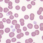
Canine
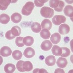
The canine erythrocyte in health is a relatively large, uniform, biconcave disc. This is reflected in the Wright’s stained blood film as a cell with an area of central pallor.
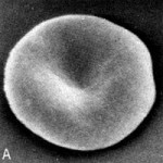
The scanning electron micrograph at the right shows this shape most graphically. This fact makes recognition of spherocytic shape changes more clear in dog blood compared to that of other species which lack significant central pallor.
Small numbers of polychromatophilic erythrocytes are observed in blood smears from healthy dogs (<1.5% reticulocytes). Occasional nucleated red blood cells and Howell-Jolly bodies may also be seen in blood smears from healthy, non-anemic dogs.
The canine erythrocyte lifespan varies from 110-120 days.
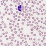
Feline
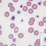
Feline erythrocytes are smaller and more variable in size and shape than those of dogs. They have little to no central pallor as normally seen in routine blood films. Polychromatophilic cells are very few in health (<1%). The hemoglobin of cats is more susceptible to oxidant injury than in other species. This is because feline hemoglobin contain eight oxidizable sulfhydryl groups. For this reason, low numbers (<10%) single, small Heinz bodies may be seen on erythrocytes in healthy cats. These so-called “endogenous” Heinz bodies may be seen in increased numbers (up to 50%) in cats on semi-moist food which contains propylene glycol that causes oxidant injury. Although these cats are not usually anemic, red blood cell lifespan is reduced. Healthy cats also can have a few Howell-Jolly bodies in erythrocytes in peripheral blood.
The lifepan of feline erythrocytes is 65-76 days. The propensity for rouleaux formation in feline erythrocytes is greater than that of canine erythrocytes, but less than that of horses.
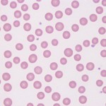
Equine
Equine erythrocytes are of similar size as feline erythrocytes and also lack central pallor. Therefore, identification of spherocytic shape change is very difficult to discern in these species. Blood from healthy horses often display prominent rouleaux formation. Occasional Howell-Jolly bodies are observed in blood smears from healthy horses. Polychromatophils are not observed in blood from healthy horses and are rarely observed in blood from anemic horses. Therefore, the degree of erythroid regeneration in response to an anemia cannot be assessed by quantifying reticulocytes in horses. The erythrocyte lifespan in horses varies from 140-150 days.
Note that microcytosis is a normal finding in foals up to 1 year of age, and is attributable to a physiologic iron deficiency (puppies and kittens can display a similar microcytosis between 4-8 weeks of age).
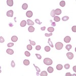
Bovine
Bovine erythrocytes are similar in size to horse erythrocytes and have a small amount of central pallor (with some lacking central pallor). Some degree of anisocytosis can be seen in smears from healthy cattle. Cattle rarely display rouleaux formation in health or disease. The approximate erythrocyte lifespan is 160 days.
Polychromatophils are not usually observed in blood smears from healthy non-anemic cattle, although reticulocytes are released along with macrocytes in response to an anemia. However, their capacity for regeneration is not as marked as the dog. Basophilic stippling of nucleated and non-nucleated erythrocytes can be a common finding in ruminants with a regenerative response.
Note that marked poikilocytosis (variation in red cell shape), thrombocytosis (often > 1 million cells/uL) and microcytosis are features of healthy calf blood (usually under 3 months of age). Calves can remain microcytic for up to 1 year of age, which is attributed to a physiologic iron deficiency.
Caprine
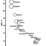
Marked poikilocytosis can be a normal feature in the blood of some goats (especially Angora) and is especially prominent in kids (< 3 months of age). Goat erythrocytes are the smallest of the domestic animal species, with mean corpuscular volume ranging from 16-25 fL. The caprine erythrocyte lifespan is approximately 125 days.
Goats do not display a prominent reticulocytosis in response to an anemia and lack polychromasia in peripheral blood in health. They do (unlike horses) release polychromatophils from the bone marrow.
Camelid
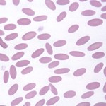
Small, flat, oval-shaped erythrocytes are typical of llamas, camels, and alpacas. The mean corpuscular hemoglobin concentration (MCHC) in camelids are significantly higher than other species, commonly running in the range of 40-50 g/dL.
A small degree of anisocytosis and poikilocytosis may be observed in blood smears from healthy llamas. The average erythrocyte lifespan in the llama is 60 days.