
Below is a summary of terms used to describe red blood cell morphologic features, the meaning of the term and associated interpretation (the provided list of diseases is not exhaustive). In addition, the interpretation is based on associations and the meaning in a given patient is entirely dependent on what is going on in the animal (i.e. context-dependent). In some cases, a cause for the change or relevance thereof, is uncertain.
| Term | Meaning | Interpretation (common or reported associations) |
Anisocytosis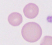 |
Variation in size (diameter) | This can be due to increased numbers of large RBC or small RBC, or a combination of both. Some degree of anisocytosis is normal in animals.This is the smear equivalent of the red blood cell distribution width (RDW), which is a measure of the variation in RBC volume. Result reporting: Subjectively graded as mild, moderate or marked. |
Acanthocytes 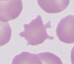 |
Irregularly spiculated RBC or “spur” cell | Mechanism: Mostly unknown, but speculated to be due to alterations in lipid composition of RBC membrane or fragmentation injury to RBCs Physiologic: Young ruminants (calves). Disorders: Cancer, including hemangiosarcoma, (canine), liver disease (canine, feline), disseminated intravascular coagulation (DIC) (canine, bovine), vasculitis (canine), iron deficiency anemia (canine, mechanical fragility), possible congenital/inherited disorder (canine), various other diseases (e.g. renal, gastrointestinal, cardiac, canine). Result reporting: Subjectively graded as few, moderate, many. Differentiate from: Echinocytes (regularly spiculated). Can be difficult in individual animals. |
Acuminocyte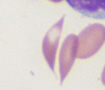 |
Fusiform RBC, spindled RBC | Mechanism: Likely polymerization of hemoglobin variants. Physiologic: Young goats, some species of adult goats (Angora), low numbers in non-anemic camelids. Diseases: By themselves, this poikilocyte does not provide much information. Increased numbers can be seen in iron deficiency anemia in camelids (unknown mechanism), increased numbers can be seen in regenerative anemia in goats. Result reporting: Subjectively graded as few, moderate, many. |
Aggregate reticulocyte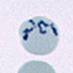 |
Aggregates of RNA in a reticulocyte stain, e.g. new methylene blue | Term used to identify immature RBC with large amounts of RNA that precipitate as large chunks or “aggregates” when the blood is incubated with an intravital dye, such as new methylene blue. Aggregate reticulocytes correspond to polychromatophilic RBC in a Romanowsky-stained blood smear (e.g. Wright’s, May-Grunwald-Giemsa, rapid stains). Relevance: Indicate a regenerative response in all species. Have a short half-life in cats (around 12 hours) so indicate the current response by the bone marrow to an anemia. May not be released from the bone marrow in mild anemias in cats. Included in a reticulocyte count in dogs and cats. |
Agglutination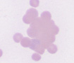 |
Clumping of RBC | Mechanism: Mediated by antibody bridging of adjacent RBC. Artifact: EDTA-dependent antibody binding. Drugs: Heparin therapy (horses). Disorders: Immune-mediated hemolytic anemia (dog, cat, horse). Result reporting: Present or absent (not graded). Differentiate from: Rouleaux formation (does not usually disperse on saline dilution). |
Basophilic stippled RBC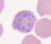 |
RBC with chunky blue dots in a Romanowsky stain | Mechanism: Aggregates of RNA due to RBC immaturity or inhibition of RNA degradation. Does not require intravital dye precipitation to observe in a regularly stained blood smear (Romanowsky stain). Physiologic: Regenerative anemia (ruminants in particular, also dogs and cats but infrequent in the latter). Pathologic: Lead poisoning (inhibits RNA degradation). Result reporting: Subjectively graded as few, moderate, many. Differentiate from: siderocytes (more focal, smaller, lighter blue dots). |
Dacryocytes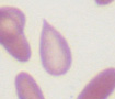 |
Tear-drop RBC | Mechanism: Unknown. Physiologic?: Low numbers in non-anemic camelids. Disorders: By themselves, this poikilocyte does not provide much information. Increased numbers can be seen in camelids with iron deficiency (mechanism unclear; perhaps due to mechanical fragility), myelofibrosis (dogs, not a consistent finding). Result reporting: Subjectively graded as few, moderate, many. |
Drepanocytes |
Sickled RBC | Mechanism: Specific types of hemoglobin form linear polymers under oxygenated states (frequently after blood sample collection). Physiologic: Deer, antelope, sheep (certain breeds) mongoose, genet. Result reporting: Subjectively graded as few, moderate, many. |
Eccentrocytes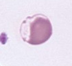 |
Hemighost with “eccentric” central pallor | Mechanism: Oxidant-induced cross-linking of RBC membrane. Drugs: Vitamin K1, phenothiazine drenches (horse), new methylene blue, propofol, acetaminophen (cats). Disorders: Copper poisoning (sheep), red maple leaf toxicity (horses, camelids), Pistacia toxicity (horses), onions (dogs, cats, cattle, horses), Brassica species, e.g. kale (ruminants), zinc toxicity (dog), naphthalene in mothballs (dog), skunk musk (dogs, panda), inherited defects (glucose-6-phosphate dehydrogenase deficiency, flavin adenine dinucleotide deficiency; both in horses), various diseases in dogs associated with oxidant injury (lymphoma, diabetes mellitus, anticoagulant rodenticide toxicosis). Result reporting: Subjectively graded as few, moderate, many. |
Echinocytes  |
Regularly spiculated,”burr” cells or crenated cells | Mechanism: Expansion of the outer leaflet of RBC membrane, ATP depletion. Artifact: Stored (aged) blood, excess EDTA, increased pH. Drugs: Furosemide (horses), salicylates, phenylbutazone, doxorubicin (small animals). Disorders: Electrolyte depletion, glomerulonephritis, pyruvate kinase deficiency, snake envenomation (rattlesnake, coral snake, viper), burns, bee stings, Clostridia infections. Result reporting: Subjectively graded as few, moderate, many. Differentiate from: Acanthocytes (irregularly spiculated). This can be difficult. |
Elliptocytes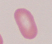 |
Oval or elongate RBC | Three types: Type I (slightly oval), type II (oval), type III (elongate). Mechanism: In inherited conditions, due to alterations in membrane (band 4.1) or cytoskeletal proteins (spectrin). Unknown mechanism in myelofibrosis. Physiologic: Camelids (low numbers). Drugs: Chemotherapeutic agents. Disorders: Myelofibrosis (dogs), hereditary disorders in spectrin and band 4.1 (dog). Result reporting: Subjectively graded as few, moderate, many. |
Ghost RBC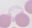 |
Lysed RBC | Mechanism: Rupture of RBC membrane, releasing hemoglobin and leaving membrane scaffolds. Artifact:In vitro hemolysis (poor sample collection, freezing of blood, aged RBC, blood smear preparation). Drugs: DMSO (horse). Diseases: Intravascular hemolytic anemia, due to immune-mediated hemolytic anemia in dogs, Babesia infection, copper poisoning in sheep, zinc toxicity in dogs, Clostridial toxins, hypophosphatemia, acute liver failure in horses, acute transfusion reaction, neonatal isoerythrolysis in horses, ruminants (not an exhaustive list). Result reporting: Subjectively graded (few, moderate, many)/ |
Heinz bodies 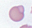 |
Refractile or red inclusions in RBC | Indicates oxidant injury. More readily visualized in a reticulocyte (new methylene blue) stain. Mechanism: Precipitated oxidized hemoglobin. Diseases: Oxidant injury (see eccentrocytes for diseases). The hemoglobin of cats is more susceptible to oxidant injury than other species, so low numbers of Heinz bodies are seen in the blood of non-anemic cats. In fact, some non-anemic cats can have many small refractile Heinz bodies due to endogenous (e.g. diabetes mellitus, hyperthyroidism, lymphoma) or exogenous oxidants (e.g. propylene glycol). These small Heinz bodies are called “endogenous” Heinz bodies. In other species, Heinz bodies are associated with an oxidant-induced hemolytic anemia. Result reporting: Subjectively graded as few, moderate, many(easier to do in a new methylene blue stain). |
Howell-Jolly body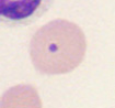 |
Nuclear remnant or micronuclei | Mechanism: Increased RBC production (regenerative anemia), decreased removal (by splenic macrophages). Physiologic: Regenerative anemia, low numbers of Howell-Jolly bodies can be seen in healthy animals (horses and cats, particularly). Drugs: Corticosteroids (inhibit splenic removal). Diseases: Dyserythropoietic syndromes (congenital, neoplasia). Result reporting: Subjectively graded as few, moderate, many. |
Hypochromic RBC  |
Less hemoglobin or hypochromasia | Mechanism: Decreased or inhibited hemoglobin production. Physiologic: Very young animals (physiologic iron deficiency anemia, easiest to identify in dogs). Mineral/nutrient deficiency: Iron deficiency (blood loss, nutritional deficiency), vitamin B6 deficiency (rare), copper deficiency (leads to iron deficiency), zinc excess. Diseases: Chronic external blood loss, chronic lead poisoning. Result reporting: Subjectively graded as mild, moderate, marked. Differentiate from: Torocytes (normal rim of hemoglobin). |
Keratocytes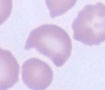 |
“Bite”, “helmet” or “blister” cells | Mechanism: Oxidant or fragmentation injury. Low numbers may be seen in non-anemic cats. Diseases: See above disorders for acanthocytes (fragmentation) and eccentrocytes (oxidant). Result reporting: Subjectively graded as few, moderate, many. |
Macrocytes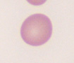 |
Bigger RBC | Not synonymous with macrocytosis (high MCV). Low numbers of macrocytes may be seen without a high MCV (insufficient numbers to increase the MCV above the upper reference limit). Mechanism: Immature RBC (larger than normal), uptake of water, altered DNA metabolism. Breed-associated: Poodles (toy and miniature), possibly Greyhounds. Artifact: Stored (aged) blood (may be associated with a low mean cell hemoglobin concentration), hyperosmolality (hypernatremia, hyperglycemia with the ADVIA hematology analyzer). Drugs: Interfere with DNA metabolism, e.g. hydroxyurea, sulfonamides. Physiologic: Response to regenerative anemia (punctate reticulocytes). Mineral/nutrient deficiency: Vitamin B12 deficiency, folate deficiency, cobalt deficiency, molybdenum excess. Diseases: Feline leukemia virus infection (cats), congenital dyserythropoietic syndromes (poll Hereford), myelodysplastic syndrome, diabetes mellitus (RBC swelling), hyperthyroidism (unknown mechanism). Result reporting: Subjectively graded as few, moderate, many. |
| Microcytes | Smaller RBC | Difficult to identify true microcytes and RBC are not usually reported as microcytes in animals with MCV below the lower reference limit for that species. This term is rarely or not used at Cornell University, since it is of uncertain relevance and could encompass more diagnostic shapes, such as spherocytes, schistocytes. Identification of the latter specific shapes is more informative. |
Poikilocytes |
Variable RBC shapes (too variable to quantify individual shapes) | Mechanism: Various (see specific changes). Physiologic: Young calves and goats, normal in adult Angora goats. Drugs: Chemotherapeutic agents, e.g. doxorubicin. Diseases: Liver disease (cats). Result reporting: Subjectively graded as few, moderate, many. |
Polychromatophils 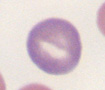 |
Purple RBC or polychromasia | Mechanism: Immature RBC which contain abundant RNA (ribosomes, polyribosomes). Correspond to aggregate reticulocytes. Physiologic: Can be seen in normal dogs and cats in low numbers. Part of a regenerative response. Result reporting: Subjectively graded as mild, moderate, marked. |
Punctate reticulocyte |
Small dots of RNA in a reticulocyte stain, e.g. new methylene blue | Term used to identify immature RBC with small amounts of RNA that precipitate as small “punctate” dots chunks when the blood is incubated with an intravital dye, such as new methylene blue. They are considered more mature than aggregate reticulocytes, because they contain less RNA. Punctate reticulocytes may be larger than normal and would correspond to macrocytes in a Romanowsky-stained blood smear (e.g. Wright’s, May-Grunwald-Giemsa, rapid stains). However, not all macrocytes are punctate reticulocytes (macrocytes can form through other mechanisms). Relevance: Included in a reticulocyte count in dogs, but not cats (can be counted separately from aggregate reticulocytes by certain laboratories; this is not done at Cornell University). Have a longer half-life in cats than aggregate reticulocytes (around 3 days) so do not indicate the current response by the bone marrow to an anemia. Only punctate reticulocytes may be released in mild anemias in cats. |
Pyknocyte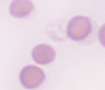 |
RBC remnant | Mechanism: Oxidant injury (remnant after removal of tags of membrane associated with rupture of eccentrocytes). Diseases: See above for eccentrocytes. Result reporting: Not always quantified or reported in hemograms. Differentiate from: Spherocytes (can only be done by electron microscopy, however usually accomplished by the company they keep, i.e. the presence of eccentrocytes would support the cells being pyknocytes versus spherocytes). |
Rouleaux formation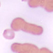 |
Stacking of RBC | Mechanism: Decreased negative charge on RBC, usually due to increased globulins (fibrinogen, immunoglobulins). Physiologic: Horses, cats, pigs (can be normal in this species). Diseases: Inflammation (high fibrinogen, polyclonal gammopathy, restricted oligoclonal gammopathy), antigenic stimulation (polyclonal or restricted oligoclonal gammopathy), neoplasia of B cells (lymphoma, chronic lymphocytic leukemia) or plasma cells (multiple myeloma, extramedullary plasmacytome, solitary myeloma of bone) producing a monoclonal immunoglobulin. Should be associated with a high total protein by refractometer or high globulin on a chemistry panel. Result reporting: Subjectively graded as mild, moderate, marked. Differentiate from: Agglutination (three-dimensional clumps): Disperses with saline dilution (1:4 to 1:10 blood:saline) |
Schistocytes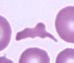 |
RBC fragments or”schizocyte” | Mechanism: Shearing of RBC in the circulation due to abnormalities in the vasculature (endothelial cell, fibrin strands, blood flow) or mechanical RBC fragility (iron deficiency). Diseases: See above for acanthocytes. Also portosystemic shunts (altered blood flow). Result reporting: Subjectively graded as few, moderate, many. |
Siderocytes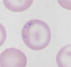 |
Aggregates of iron, focal blue/gray inclusions, siderotic granules, pappenheimer bodies | Mechanisms: Aggregates of iron (in lysosomes or mitochondria), due to increased iron turnover or inhibition of iron use. Drugs: Hydroxyzine, chloramphenical. Diseases: Hemolytic anemia (increased RBC turnover), myelodysplastic syndrome and acute myeloid leukemia (cats), lead poisoning, portosystemic shunts, vitamin B6 and copper deficiency in pigs. Result reporting: Subjectively graded as few, moderate, many. Differentiate from: Basophilic stippling (coarse chunks of RNA, diffusely distributed around RBC): Would stain positive for iron with a Prussian blue stain. |
Spherocytes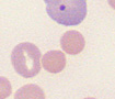 |
Sphered RBC | Mechanism: Removal of membrane by macrophages (trogocytosis). Artifact: Stored red blood cells (transfusions), feathered edge. Disease: Immune-mediated hemolytic anemia (primary or secondary to infectious agents, drugs), pyruvate kinase deficiency (spheroechinocytes), fragmentation (low numbers), hemophagocytic syndrome, histiocytic sarcoma, snake envenomation, inherited band 3 deficiency in Japanese black cattle. Result reporting: Subjectively graded as few, moderate, many. Differentiate from: Pyknocytes (context-dependent, the company they keep, electron microscopy) or microcytes (can be difficult if only a few but latter are more variable in size). |
Stomatocytes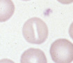 |
RBC with a slit or mouth-like central pallor (“stoma”) | Mechanism: Expansion of the inner leaflet of the RBC membrane. Artifact: Blood smear preparation. Physiologic: Woodchuck, manatee, dolphin. Disorders: Hereditary stomatocytosis in dogs (Alaskan Malamute, Drentje patrishond, standard and miniature Schnauzer, Peek-a-poo, Pomeranian). Result reporting: Subjectively graded as few, moderate, many. |
Target cell  |
RBC with a bullseye. Codocyte | Only recognized in dogs, which have central pallor. Mechanism: Expansion of the inner leaflet of the RBC membrane from alterations in phospholipid:cholesterol content in the membrane, cells that spread in a smear than normal (leptocytes) because they are larger than normal (polychromatophils) or thinner than normal (iron deficient cells). Diseases: Iron deficiency anemia (hypochromic RBC), alterations in lipid content occurs in liver disease or dyslipidemias. Result reporting: Subjectively graded as few, moderate, many. |
Torocyte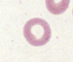 |
Punched out cell | Artifact: Blood smear preparation. Result reporting: Not usually reported on a hemogram. Differentiate from: Hypochromasia (normal rim of hemoglobin, “punched out” central pallor with sharp edges). |