
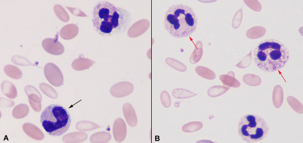
These images are from an iron deficient alpaca that also had an inflammatory leukogram due to an enteritis.
A:The two cells are a segmented neutrophil (upper cell) and a band neutrophil (black arrow). Toxic change is evident in both cells – cytoplasmic vacuolation and toxic granulation in the segmented neutrophil and cytoplasmic basophilia in the band neutrophil.
B:Three segmented neutrophils are shown, all of which are toxic, demonstrating toxic granulation (upper two cells identified with the red arrows) and cytoplasmic vacuolation (all three cells). The red blood cells in the background are hypochromic (have less hemoglobin than normal) which is a cardinal feature of iron deficiency.