
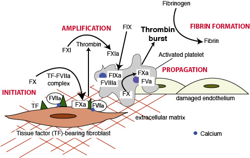
In the cell-based model, thrombin generation and fibrin formation proceeds on cell surfaces. In the initiation phase of thrombin generation, plasma FVII binds to tissue factor (TF) on subendothelial fibroblasts. Once bound to TF, FVII autoactivates and the TF-FVII complex forms the “extrinsic tenase”, which activates FX on fibroblast membranes. The extrinsic pathway is rapidly inhibited by tissue factor pathway inhibitor and produces only a small amount of thrombin. The small amount of thrombin generated by the extrinsic pathway, then amplifies its own production on phosphatidylserine (PS)-rich surfaces of activated platelets, by activating FXI of the intrinsic pathway and FV and FVIII, the intrinsic and common pathway cofactors. FXIa then activates FIX, which forms a potent “intrinsic tenase” complex with FVIIIa on the platelet surface. The PS-enriched surfaces of activated platelets helps amplify and propagate thrombin generation, producing a “thrombin burst”. The thrombin burst is essential for forming crosslinked fibrin from fibrinogen, by cleaving fibrinogen to soluble fibrin, activating factor XIII, which crosslinks fibrin, while concurrently inhibiting fibrinolysis. The formed fibrin closely intercalates and binds to the platelet plug forming a stable fibrin clot.
Note that calcium is crucial for all aspects of fibrin formation and allows coagulation factors to bind to membranes.
“Reproduced and modified with permission from the BSAVA Manual of Canine and Feline Clinical Pathology, 3rd edition.”