
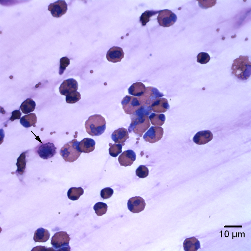
Figure 2a: Eosinophils dominate (with some free eosinophil granules) with low numbers of lightly granulated mast cells (arrow) (Wright’s stain, 50x objective).
Figure 2a: Eosinophils dominate (with some free eosinophil granules) with low numbers of lightly granulated mast cells (arrow) (Wright’s stain, 50x objective).
eClinpath helped 1.2 million visitors last year from 220 countries find important information on animal health. If you enjoy the site, please support our mission and consider a small gift to help us keep pace with its rapid growth. You can donate securely via PayPal or credit card. Thank you!
![]()