
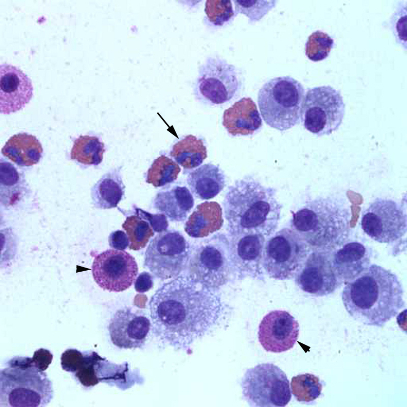
The bronchoalveolar fluid contained a mixed population of cells, which were mostly macrophages and eosinophils (38%, arrows), with increased proportions of mast cells (10%, arrowhead). Some erythrocytes are present in the background (Wright’s stain, 50x objective)