
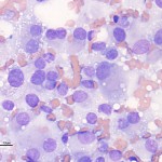
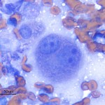
Photomicrographs of an aspirate from a subcutaneous mass from a dog
Case information
A 9 year old castrated male Border Collie was presented for a chief complaint of a rapidly growing subcutaneous mass over the left shoulder. The mass had doubled in size over a period of three months; this increase in size coincided with progressive left forelimb lameness.
On presentation, the dog was not bearing weight on the left forelimb. A firm mass, approximately 5 inches in diameter, was palpable over the left shoulder and appeared to be continuing into the axillary region. Muscle atrophy was observed on the left forelimb and both hindlimbs. Pain was elicited on palpation of the left axillary region. The dog resented manipulation of the left shoulder, though the range of motion in the left forelimb was adeqaute. Thoracic auscultation revealed a grade 4/6 systolic heart murmur and normal lung sounds in all fields. No abnormalities were appreciated on abdominal palpation or rectal examination.
Abdominal ultrasound, thoracic radiographs, and a hemogram revealed no relevant abnormalities. The mass did not appear to be involving bones in the shoulder region. The serum chemistry panel showed a mild increase in creatinine (1.7 mg/dL, reference interval, 0.6-1.4 mg/dL), a moderate hypercalcemia (14.8 mg/dL, reference interval, 9.3-11.4 mg/dL), with a high ionized calcium of 1.66 mmol/L (reference interval 1.16-1.40 mmol/L), and a mildly increased alkaline phosphatase (210 U/L, reference interval, 17-111 U/L). Urinalysis (voided) revealed 30 mg/dL (1+) protein with a urine specific gravity of 1.018. The urine sediment was inactive (no evidence of hematuria, pyuria or bacteriuria). A fine needle aspirate was performed on the subcutaneous mass. See the representative images of a Wright’s stained smear of the aspirate of the shoulder mass, then answer the questions below:
- Based on the cytologic features of the cells, what is your differential diagnosis for the mass?
- What additional diagnostic tests could be performed on the cytology smears to facilitate a definitive diagnosis?
- Is the hypercalcemia related to the subcutaneous mass?
|
|
|
Answer on next page