
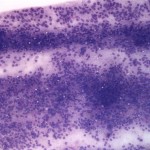
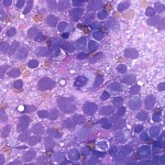
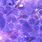
Smear from a biopsy of a brain mass in a dog
Case information
A 4 year old female spayed boxer was presented for a 2 day history of cluster seizures. The owner reported that the seizures first started as multiple focal facial seizures, including foaming at the mouth and chomping. On the day of presentation, the dog had also suffered from additional multiple episodes of generalized seizures, which lasted approximately 15-20 seconds. Upon presentation, the dog was bright and alert. No abnormalities were detected on physical examination and results of a hemogram and chemistry were unremarkable. The dog was transferred to the section of neurology in the Cornell University Hospital for Animals. A neurologic examination localized the lesion to the forebrain. The dog was started on anticonvulsant therapy with a loading dose of zonisamide (250mg every 12 hours) and pulse therapy of Keppra (250mg every 8 hours). Magnetic resonance imaging revealed a focal, intra-axial, minimally contrast-enhancing mass in the right piriform lobe with mild deviation of normal parenchymal tissue. Analysis of cerebrospinal fluid revealed no cytologic abnormalities. The dog was taken to surgery and smears were made from a surgical biopsy of the mass (Figure 1-3). After examining the photomicrographs of the smears of the mass, answer the questions below:
- What disease process is evident in the aspirate from the brain mass?
- What are the differential diagnoses for a brain mass in the dog?
|
|
|
|
Answer on next page