
Crystalluria in a Dog
Case information
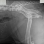
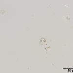
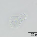
A 1-year-old intact male English Mastiff was presented to Emergency Medicine at the Cornell University Hospital with a one week history of pollakiuria and stranguria. Four days prior to presentation, the dog had been started on oral prazocin (an α-1 adrenergic blocker) with no improvement. Hematuria, coughing, sneezing, or diarrhea were not reported. The patient had vomited food material and was inappetent in the 2 days prior to presentation. On physical examination, the patient was 5% dehydrated and was noted to be arching the lumbar spine. No abnormalities were noted on abdominal palpation. No abnormalities were detected on a hemogram and mild electrolyte changes (sodium 154 mEq/L, reference interval, 143-150 mEq/L; chloride 115 mEq/L, reference interval, 106-114 mEq/L) were evident on a biochemical panel. Abdominal radiographs were also obtained (Figure 1). Urine collected by cystocentesis was medium yellow, slightly cloudy with a urine specific gravity of 1.014, pH of 8.5, and trace protein on a dipstick. On urine sediment examination, moderate numbers of sperm and the crystals pictured below (Figures 2-3) were seen.
Evaluate the provided cytologic images (Figures 1-3) and answer the following questions:
- What differential diagnosis would you have for this patient based on the history and imaging results?
- What is your main differential diagnosis based on the crystalluria and what is unusual about the urinalysis in this case?
- What additional treatment and tests would you recommend?
|
|
|
|
Answers on next page.