
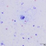
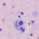
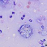
Subretinal aspirate from a dog
Case Information
A 7 year old castrated male mixed breed dog presented with a 9 month history of intermittent watery, mucoid and bloody diarrhea, which was not completely responsive to medical therapy and a hypoallergenic prescription diet. The patient subsequently developed panuveitis and was referred to Cornell University Hospital for Animals after starting treatment with prednisolone acetate eye drops and oral prednisone (5mg twice daily). Upon initial presentation, most of the physical examination was unremarkable, except there was still bilateral panuveitis, with 50 and 100% retinal detachment in the right and left eyes, respectively. A subretinal aspirate was performed of the left eye and submitted for cytologic evaluation.
Evaluate the provided cytologic images (Figures 1-3) and answer the following questions:
- What differential diagnoses would you have for this patient based on the history?
- What are the rod-like structures within the macrophages and scattered in the background?
- What additional tests would you recommend?
|
|
|
|
Answers on the next page